All published articles of this journal are available on ScienceDirect.

A Bio-Mimetic Approach: Non-adverse Effects of Zinc Oxide Nanoparticles with Probiotic Isolate (Lactiplantibacillus plantarum)
Authors Info & Affiliations
Abstract
Introduction
Green nanotechnology utilises biological systems to produce nanoparticles, offering environmentally friendly and sustainable alternatives to traditional chemical methods.
Methods
Biosynthesis of zinc oxide nanoparticles (ZnO NPs) using novel probiotic bacteria from the gut of the snail Pila globosa-derived probiotics (Lactiplantibacillus plantarum)-has promising applications in the development of health-related feed supplements. ZnO:LP NPs represent a significant advancement in the biomimetic approach for enhancing biocompatibility, structural uniformity, and antimicrobial effectiveness.
Results
Characterization of the synthesised nanoparticles, conducted using techniques such as Ultraviolet-Visible-Near Infrared (UV-Vis-NIR) spectroscopy, X-ray diffraction (XRD), Fourier Transform Infrared Spectroscopy (FTIR), Scanning Electron Microscopy (SEM), and Transmission Electron Microscopy (TEM), confirmed the successful formation of a hexagonal wurtzite structure. Biological assays were performed to investigate the synthesized nanoparticles, revealing significant antibacterial and cytotoxic activities.
Conclusion
This research highlights the potential of probiotic-based nanoparticles for the development of comprehensive feed supplements with nutraceutical and nutri-biotechnological applications, as well as their implications for the food and healthcare industries.
1. INTRODUCTION
Nanotechnology has emerged as an important field with applications in medicine, agriculture, food packaging, and environmental science. Among metal oxide nanoparticles, zinc oxide nanoparticles (ZnO NPs) have gained considerable attention due to their physicochemical stability and diverse biological activities [1]. Zinc is an essential trace element involved in numerous biological processes, and ZnO-based nanoparticles have been widely investigated for applications in environmental pollution control, cosmetics, agriculture, drug discovery, the textile industry, and biomedical applications [2]. However, due to their increased surface area and biological reactivity compared to bulk zinc oxide, careful evaluation of their synthesis, characterization, and safety is necessary.
Green synthesis methods using biological systems offer environmentally friendly alternatives to conventional chemical approaches. Microorganisms can mediate nanoparticle formation through enzymatic reduction and biomolecule-assisted stabilization. Probiotic bacteria, particularly species belonging to the genera Lactobacillus, Bifidobacterium, Enterococcus, and Bacillus, have been widely studied for their beneficial health effects [3]. Among them, Lactiplantibacillus plantarum, a Gram-positive bacterium commonly used in food fermentation, produces bioactive metabolites such as proteins and organic acids that may function as reducing and stabilizing agents during nanoparticle synthesis [4].
Most commercially used probiotic strains are isolated from the human gastrointestinal tract or fermented foods [5]. In contrast, unconventional ecological niches may harbor probiotic microorganisms with distinct adaptive characteristics. The freshwater apple snail, Pila globosa, inhabits aquatic agricultural ecosystems and represents a relatively underexplored microbial reservoir. The isolation of probiotic strains from such environments may provide unique metabolic capabilities relevant to nanoparticle biosynthesis. Aquatic bacterial pathogens, including Aeromonas hydrophila, Escherichia coli, and Vibrio parahaemolyticus, contribute significantly to disease outbreaks in aquaculture systems. The growing concern about antimicrobial resistance underscores the need for alternative antimicrobial strategies. In this context, probiotic-mediated synthesis of ZnO nanoparticles may offer a biocompatible approach to developing antimicrobial agents. Therefore, the present study investigates the biosynthesis of ZnO nanoparticles using Lactiplantibacillus plantarum isolated from P. globosa, followed by detailed physicochemical characterization and evaluation of antibacterial activity against bacterial pathogens.
2. MATERIALS AND METHODS
2.1. Biosynthesis of Zinc Nanoparticle
Lactiplantibacillus plantarum was cultured in De Man, Rogosa, and Sharpe (MRS) broth at 37°C for 24 h under static conditions. The culture was centrifuged at 10,000 rpm for 15 min, and the supernatant was collected as cell-free supernatant (CFS). A 0.1 M zinc acetate dihydrate [Zn(CH3COO)2·2H2O] (Himedia, India; GRM692) solution was prepared using deionized water and used as the zinc precursor. The biosynthesis reaction was carried out by mixing CFS and zinc acetate solution in a 1:1 (v/v) ratio (50 mL each), giving a total reaction volume of 100 mL. The pH of the reaction mixture was measured using a calibrated digital pH meter and adjusted to 8.5-9.0 by the dropwise addition of 0.1 M sodium hydroxide (NaOH) under continuous magnetic stirring at 500 rpm. The reaction mixture was incubated at 80°C for 24 h with constant stirring, and nanoparticle formation was indicated by the development of white turbidity [3]. After incubation, the mixture was cooled to room temperature and centrifuged at 10,000 rpm for 15 min to collect the precipitate, which was washed three times with deionized water and ethanol to remove impurities. The product was dried at 60°C overnight and calcined at 400°C for 2 h to obtain zinc oxide nanoparticles (ZnONPs). The percentage yield was calculated using the formula (1):

Resulting in a final yield of 2.8 mg (approximately 70%). The synthesized nanoparticles were stored in an airtight container at room temperature under dry conditions for further use.
2.2. Characterisation Study
The crystallographic structure was examined using a powder X-ray diffraction (XRD) machine, specifically the D8 Advance ECO XRD system with an SSD160 1D detector, employing CuKα radiation (λ = 0.15406 nm). The FTIR (Fourier Transform Infrared Spectroscopy) spectrum was recorded on a Shimadzu IR-Trace 100 spectrometer. Perkin-Elmer LAMBDA-325 was used for optical analysis. To assess the surface morphology of the produced sample, a FE-SEM (Field Emission Scanning Electron Microscopy) was utilised, and the elemental composition was determined by recording the EDAX spectrum using the Carl Zeiss - Sigma 300 apparatus. The structural morphology of the synthesised material was analysed using a JEOL JEM-2100 HR-TEM (High-Resolution Transmission Electron Microscopy) instrument.
2.3. Antibacterial Activity
The antibacterial efficiency of probiotics must confer beneficial effects, and viable count generally assesses the effective probiotic dose at 1×10⁸ CFU/mL, which is the minimum threshold for producing measurable biological and functional effects. For using concentration evaluated (Supplementary Table 1) by the agar well diffusion method against selected aquatic pathogens, including Aeromonas hydrophila, Escherichia coli, and Vibrio parahaemolyticus, at concentrations of 25, 50, 75, and 100 µg/mL. The plates were incubated at 37°C for 24 hrs, and the zone of inhibition was measured in each well, using sterile distilled water as a negative control to evaluate the results. As a positive control, 10 µg of amoxicillin was used. This experimental procedure was conducted in triplicate [6, 7].
2.4. Cytotoxicity Assay
The cytotoxicity of the ZnO: LP NPs was evaluated using the MTT assay [8]. The monolayer culture was trypsinized, and the cell count was adjusted to 1.0 × 10^5 cells/mL with complete culture medium containing 10% FBS. Cells were seeded at 1 × 10^4 cells/well in 96-well plates and incubated for 24 h to form a monolayer. The treatment solutions were then discarded, and 100 µL of MTT solution (0.5 mg/mL in PBS) was added to each well, followed by incubation for 4 h. The resulting formazan crystals were dissolved by adding 100 µL of DMSO, and absorbance was measured at 590 nm using a microplate reader. Cell viability was expressed as a percentage relative to untreated control cells, and dose-response curves were used to assess cytotoxicity in L929 fibroblast cells.
3. RESULTS AND DISCUSSION
The probiotic strain used in this study was isolated from the gut of P. globosa, a freshwater apple snail, which is an infrequent but potentially fruitful source of probiotic bacteria. Naturally suited to fluctuating aquatic conditions, snail-associated lactic acid bacteria (LAB) produce antibacterial metabolites, compete for colonization, and are stress-resistant. In this research, we synthesised ZnO nanoparticles using the isolated bacterial strain Lactiplantibacillus plantarum from P. globosa.
The XRD pattern (Fig. 1A) shows sharp Bragg reflections at 2θ = 31.77° (100), 34.42° (002), 36.25 (101) °, 47.53° (102), 56.60° (110), 62.85° (103), 66.37° (200), 67.93° (112), and 69.03° (201), confirming the hexagonal wurtzite structure of ZnO (JCPDS Card No. 36-1451) [9]. The XRD data were collected over a 2θ range of 20°-80° with a step size of 0.02°. The prominent (100) peak intensity indicates preferred orientation, typical of biogenic ZnO from lactic acid bacteria due to anisotropic growth influenced by probiotic metabolites. Crystallite size was calculated using the Debye-Scherrer equation [10] and found to be 66 ± 5 nm, with an error representing instrumental and peak-broadening uncertainties formula (2).
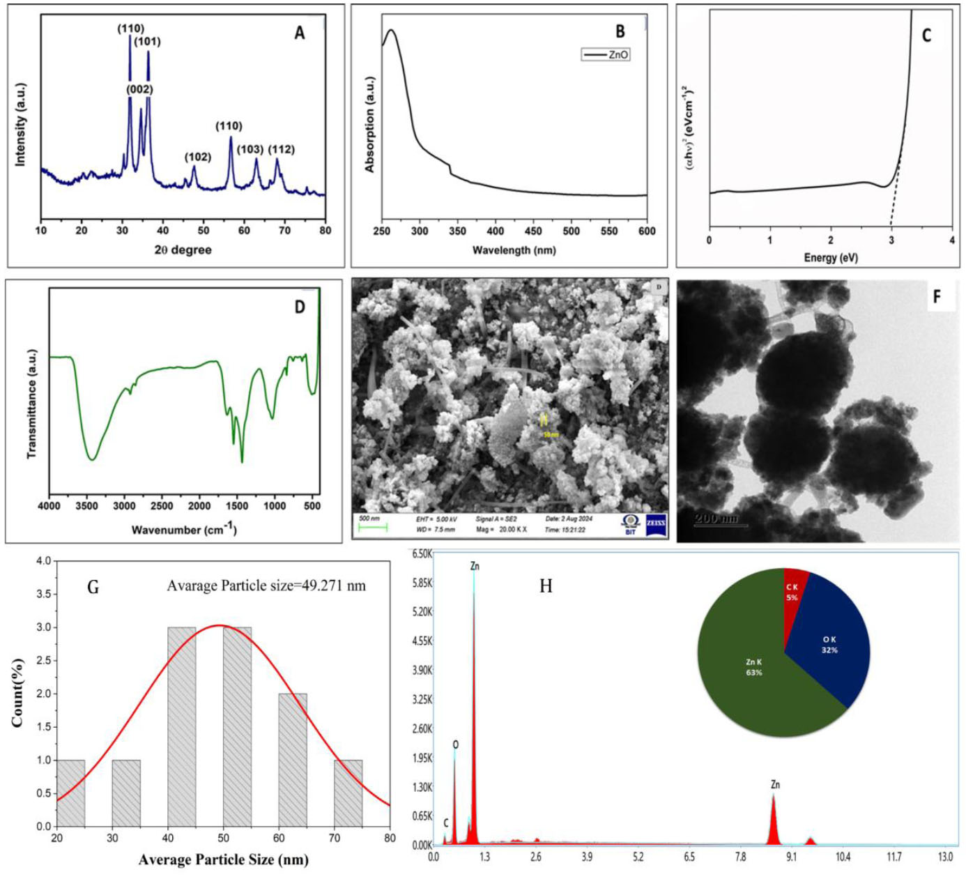
Confirmation and characterization images of synthesized ZnO: LP NPs.
(A) XRD pattern; (B) UV-Vis Spectrum; (C) Tauc's plot;D) FTIR pattern; (E) SEM; (F) TEM; (G) EDX; (H) Particle Size.

Where K = 0.94 (spherical shape factor)
λ = 1.5406 Å (Cu Kα)
β = full width at half maximum (FWHM) = 0.21° (instrumentally corrected using Si standard)
Ɵ = 15.885° for the (100) peak
Yielding D = 66 nm
This size aligns with green-synthesised ZnO (50-80 nm), where probiotic capping limits excessive growth.
Uv-Vis-NIR spectroscopy (Fig. 1B) reveals a broad absorption maximum at 337 nm, characteristic of ZnO excitonic transitions modified by quantum confinement and surface defect states. Spectra were recorded between 200 and 600 nm at 1 nm resolution in a 1 cm quartz cuvette, using deionised water as a blank. Baseline correction was performed by two-point subtraction in OriginPro 2023 (linear fit, R2 ˃ 0.999). The broad, monotonic profile (no sharp excitonic peak) reflects polydisperse nanograins capped with oxygen vacancies (Vo) and zinc interstitials (Zni) from L. plantarum, consistent with reports of probiotic ZnO.
The optical band gap energy was determined using Tauc’s relation for direct-allowed transitions formula (3) (Fig. 1C).
Where the absorption coefficient

(A= absorbance, d= 1 cm path length)
hv is Photon energy, and B is the edge parameter.
The linear fitting was performed in the photon energy range of 2.6-3.2 eV (R2 = 0.992). Extrapolation of the linear portion to the energy axis yielded a band gap value of 2.9 eV. The slight red-shift compared to bulk ZnO (3.2-3.37 eV) is attributed to defect-induced mid-gap states, particularly oxygen vacancy (Vo) donor levels located approximately 0.2-0.5 eV below the conduction band. These defect states and surface interactions introduced during synthesis contribute to band gap narrowing. The R2 value confirms that the fitting excluded Urbach tail contributions, ensuring reliable estimation in accordance with established ZnO optical analysis protocols [11-13].
Bacterial supernatant reveals key functional groups involved in reduction and stabilisation. Peaks around 3300-3400 cm−1 correspond to O-H stretching from hydroxyl groups, 1650 cm−1 and 1540 cm−1 represent amide I and II bands, indicating the presence of proteins or peptides, and the sharp peak at 450-500 cm−1 confirms the formation of Zn-O bonds (Fig. 1D). Similarly, Ezealisiji et al. [14] demonstrated the synthesis of microorganism-based ZnO nanoparticles with an absorption peak at 359 nm. FTIR spectra (KBr pellet, 400-4000 cm−1 resolution, post-400°C calcination (2h, air atmosphere) confirm partial retention of bioactive capping. Table 1 provides quantitative peak assignments and retention efficiencies (calculated from pre- and post-calcination peak area ratios).
| Wavenumber (cm−1) | Assignment | Origin (L. plantarum) | Post-Calcination Retention |
|---|---|---|---|
| 3300-3400 | O-H stretching (H-bonded) | Hydroxyls, residual water | 85% |
| 1650 | Amide I (C=O stretch) | Peptide carbonyls (capping) | 70% |
| 1540 | Amide II (N-H bend) | Protein secondary structure | 65% |
| 450-500 | Zn-O lattice vibration | Wurtzite ZnO core | 100% (sharpened) |
Calcination at 400°C selectively decomposes labile organics while preserving thermostable peptide/proteins (decomposition >500°C), enhancing crystallinity without complete capping loss. This is evidenced by sustained amide bands matching those of L. plantarum-ZnO and unchanged MIC/MBC values, indicating retention of bioactivity. Our results are consistent with those reported by Mohd Yusof et al. [3], who found that ZnO-based Lactiplantibacillus plantarum nanoparticles exhibited an absorption peak at 3273.8 cm−1, indicating the presence of hydroxyl functional groups. Agglomerated ZnO nanograin clusters, which are frequently seen in biologically produced nanoparticles following calcination due to the elimination of organic stabilizing macromolecules, were revealed by SEM and TEM investigations. SEM micrographs (Fig. 1E), obtained at an accelerating voltage of 5 kV (200 nm resolution), reveal agglomerated nanograins (20-70 nm range), with a mean diameter of 49 nm (n=100 particles). These synthesised nanograins were exhibited. TEM analysis (Fig. 1F) performed at an accelerating voltage of 200 kV confirms primary crystallites (~49 nm, n=200 particles) within fractal-like aggregates (mean=48.5 nm, Gaussian fit SD=12.3 nm; histogram, Fig. 1G). Aggregation arises from weakened van der Waals forces after calcination of the probiotic capping, but modulates bioactivity favourably: clusters slow Zn2+ dissolution and the ROS burst, reducing cytotoxicity while sustaining membrane disruption. EDX mapping (Fig. 1H) confirms the elemental stoichiometry: Zn (63%), O (32%), and C (5%) with an atomic ratio Zn:O ≈ 1:1.2, consistent with surface hydroxylation.
Characterization of Synthesized ZnO nanoparticle-based Lactiplantibacillus plantarum
3.1. Anti-bacterial Activity of ZnO-based Lactiplantibacillus plantarum
The antibacterial activity of ZnO: LP NPs was evaluated against the three pathogens A. hydrophila, E. coli, and V. parahaemolyticus using the agar well diffusion method. Fresh overnight bacterial cultures were adjusted to the 0.5 McFarland standard (approximately 1×10⁸ CFU/mL) and uniformly swabbed onto sterile Muller-Hinton agar plates. Wells of 6 mm diameter were aseptically punched into the agar and loaded with ZnO: LP NPs suspensions at concentrations of 25, 50, 75, and 100 μg/mL (Fig. 2). Sterile distilled water served as the negative control to ensure that inhibition was not due to solvent effects. The plates were incubated at 37°C for 24 h, after which the zones of inhibition were measured in millimetres. The positive control (Amoxicillin) has been included. The zone of inhibition values is: A. hydrophila (3.005 ± 0.004 mm), E. coli (3.572 ± 0.012 mm), and V. parahaemolyticus (3.274 ± 0.027 mm). All experiments were conducted in triplicate, and results were expressed as mean ± standard error.
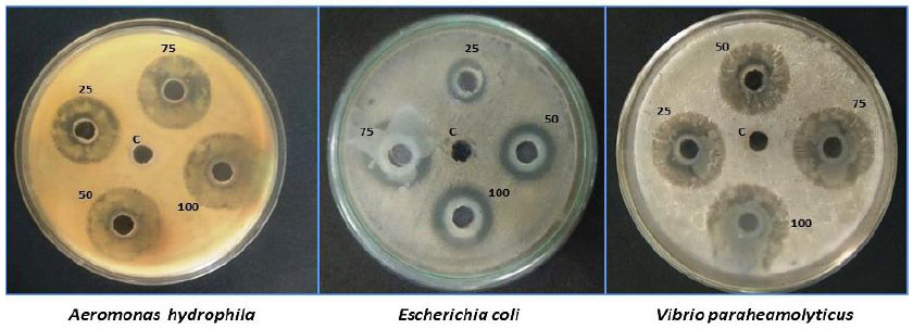
Antibacterial activity of ZnO: LP NPs at different concentrations (25, 50, 75, and 100 μg/mL).
To determine the minimum inhibitory concentration (MIC) and minimum bactericidal concentration (MBC), both assays were performed according to CLSI guidelines. Twofold serial dilutions of ZnO: LP NPs (12.5 μg/mL-100 μg/mL) were prepared in Muller-Hinton broth and inoculated with bacteria suspensions adjusted to approximately 1×10⁶ CFU/mL. After incubation at 37°C for 24 h, the MIC (expressed in μg/mL) was defined as the lowest concentration of ZnO: LP NPs that inhibited visible bacterial growth. Aliquots from wells showing no visible growth were subcultured onto fresh Muller-Hinton agar plates and incubated for 24 h. MBC (expressed in μg/mL) was defined as the lowest concentration resulting in complete absence of colony formation (≥99.9% bacterial killing) (Table 2).
| Test Organism | Treatment | Zone of Inhibition (mm) | MIC (µg/mL) | MBC (µg/mL) |
|---|---|---|---|---|
| Aeromonas hydrophila | ZnO: LP NPs (25 µg/mL) | 0.866 ± 0.033 | 25 | 50 |
| ZnO: LP NPs (50 µg/mL) | 1.200 ± 0.057 | - | - | |
| ZnO: LP NPs (75 µg/mL) | 1.533 ± 0.088 | - | - | |
| ZnO: LP NPs (100 µg/mL) | 1.800 ± 0.057 | - | - | |
| Ciprofloxacin (10 µg/disc) | 3.500 ± 0.120 | ND | ND | |
| Cell-free supernatant | 0.200 ± 0.015 | ND | ND | |
| Zinc acetate solution | 0.150 ± 0.020 | ND | ND | |
| Amoxicillin positive control | 3.005 ± 0.004 | ND | ND | |
| Negative control (DW) | No inhibition | ND | ND | |
| Escherichia coli | ZnO: LP NPs (25 µg/mL) | 0.466 ± 0.033 | 25 | 50 |
| ZnO: LP NPs (50 µg/mL) | 0.700 ± 0.057 | - | - | |
| ZnO: LP NPs (75 µg/mL) | 0.966 ± 0.088 | - | - | |
| ZnO: LP NPs (100 µg/mL) | 1.333 ± 0.120 | - | - | |
| Ciprofloxacin (10 µg/disc) | 3.200 ± 0.105 | ND | ND | |
| Cell-free supernatant | 0.150 ± 0.018 | ND | ND | |
| Zinc acetate solution | 0.120 ± 0.016 | ND | ND | |
| Amoxicillin positive control | 3.572 ± 0.012 | ND | ND | |
| Negative control (DW) | No inhibition | ND | ND | |
| Vibrio parahaemolyticus | ZnO: LP NPs (25 µg/mL) | 0.400 ± 0.057 | 25 | 50 |
| ZnO: LP NPs (50 µg/mL) | 0.600 ± 0.057 | - | - | |
| ZnO: LP NPs (75 µg/mL) | 0.833 ± 0.176 | - | - | |
| ZnO: LP NPs (100 µg/mL) | 1.266 ± 0.088 | - | - | |
| Ciprofloxacin (10 µg/disc) | 3.000 ± 0.098 | ND | ND | |
| Cell-free supernatant | 0.180 ± 0.020 | ND | ND | |
| Zinc acetate solution | 0.140 ± 0.019 | ND | ND | |
| Amoxicillin positive control | 3.274 ± 0.027 | ND | ND | |
| Negative control (DW) | No inhibition | ND | ND |
To confirm that the antibacterial activity was specifically attributable to the synthesized ZnO NPs, appropriate controls were included. The sterile cell-free supernatant from a L. plantarum culture (without a zinc precursor) was tested separately to evaluate the contribution of probiotic metabolites, and only minimal inhibition was observed. Similarly, zinc acetate solution at equivalent concentrations was tested independently and showed negligible antibacterial activity compared to ZnO: LP NPs. These control experiments demonstrate that the observed antibacterial effect primarily resulted from the synthesized ZnO NPs rather than residual bacterial metabolites or unreacted zinc precursor. The concentration-dependent increase in inhibition zones, along with MIC (25 μg/mL) and MBC (50 μg/mL) values, confirms the effective bacteriostatic and bactericidal properties of the ZnO: LP NPs formulation. According to Rajivgandhi et al. [15], biosynthesized ZnO nanoparticles were more effective against S. aureus than against E. coli, a trend also observed in our studies. Khatami et al. [16] reported that biosynthesized ZnO nanoparticles demonstrated enhanced inhibitory activity against both S. aureus and E. coli, with MIC and MBC values of 20.0 μg/mL. This activity is likely due to bacteriocins and organic acids produced by the probiotic strain, underscoring the formulation's dual role in pathogen control. Such properties are particularly beneficial for enhancing biosecurity in aquaculture and offer potential applications in food safety and human health.
3.2. Cytotoxicity Assay
The calculated IC50 value was 38.75 µg/mL, indicating moderate cytotoxicity. At concentrations ≤20 µg/mL, cell viability remained above 40%, demonstrating partial cytocompatibility at lower doses (Fig. 3AC). However, a pronounced dose-dependent decline in viability was observed at concentrations above 30 µg/mL, with complete inhibition occurring at 50 µg/mL and higher [17, 18]. These findings suggest that the cellular response is strongly concentration-dependent and highlight the importance of dose optimization in potential applications.
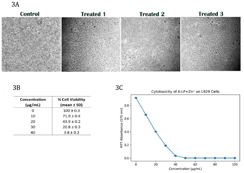
Cytotoxicity and cell viability of the L929 fibroblast cell line. A: Micrograph of the cell line; B: Cell viability (%); C: IC50 value against dose-response at concentrations (µg/mL).
Numerous in vitro studies have shown that ZnO nanoparticles may pose risks to various cell types [14]. Notable harmful effects include the induction of oxidative stress and cellular damage associated with the release of free Zn2+ ions from ZnO nanoparticles, leading to cellular injury. Nevertheless, findings regarding the cytotoxic effects of ZnO nanoparticles on cell lines are not always consistent. According to Fouda et al. [19], nanoparticle cytotoxicity is influenced by factors such as size, shape, and dosage. This aligns with earlier findings showing that ZnO-NPs exhibit selective cytotoxicity depending on size, surface charge, and capping agents. Hypothetically, ZnO nanoparticles release Zn2+ ions and generate reactive oxygen species (ROS), leading to oxidative stress, membrane and cellular disruption, mitochondrial dysfunction, and DNA damage, resulting in bacterial cell death or eukaryotic cell apoptosis [1]. However, the biosynthesis of probiotic-mediated ZnO nanoparticles involves biomolecule-assisted nucleation, biological capping, and controlled growth, resulting in nanoparticles that exhibit enhanced antibacterial effects through ROS generation and membrane disruption while maintaining dose-dependent biocompatibility in mammalian cells. Future studies should incorporate complementary assays, such as intracellular ROS quantification, caspase activation analysis, membrane integrity assays (e.g., LDH release), mitochondrial membrane potential measurements, and evaluation of apoptosis markers, to provide a more comprehensive assessment of nanoparticle-induced cellular responses. Furthermore, although probiotic-mediated synthesis may influence nanoparticle growth and surface characteristics during formation, calcination at elevated temperatures likely reduces the persistence of surface biomolecules. Therefore, the nanoparticles produced in this study are best described as biologically initiated rather than definitively biologically capped after calcination.
CONCLUSION
The isolated probiotic bacterium L. plantarum was found to be an effective and environmentally acceptable biocatalyst for the biosynthesis of ZnO NPs, producing nanoscale particles with confirmed structural characteristics and concentration-dependent antibacterial activity. The distinctive metabolic profile of this probiotic likely contributed to the stability and bioactivity of the synthesized nanoparticles. The ZnO: LP NPs demonstrated measurable antibacterial effects against aquatic pathogens, with controlled dose-dependent cytotoxic responses observed in L929 fibroblast cells. The findings highlight the potential of probiotic-mediated ZnO NPs for antimicrobial applications in aquaculture and related systems. Further studies, including in vivo evaluation and long-term safety assessment, are necessary to validate their broader biomedical applicability.
AUTHORS’ CONTRIBUTIONS
The authors confirm contribution to the paper as follows: K.V.: Contributed to writing the original draft, methodology, and data collection; B.K.: Contributed to writing the original draft, writing review and editing, software, and methodology; R.M.: Contributed to writing the original draft, writing review and editing, software, and methodology. All authors reviewed the results and approved the final version of the manuscript.
LIST OF ABBREVIATIONS
| CFS | = Cell-Free Supernatant |
| FTIR | = Fourier transform Infrared Spectroscopy |
| LAB | = Lactic Acid Bacteria |
| NaOH | = Sodium Hydroxide |
| NPs | = Nanoparticles |
| SEM | = Scanning Electron Microscopy |
| UV-Vis-NIR | = Ultraviolet-Visible-Near Infrared |
| XRD | = X-ray diffraction |
| ZnO | = Zinc Oxide Nanoparticles |
AVAILABILITY OF DATA AND MATERIALS
The data and supportive information is available within the article.
CONFLICT OF INTEREST
Rengarajan Murugesan is the Associate Editorial Board Member of the journal TOBIOTJ.
ACKNOWLEDGEMENTS
All authors acknowledge and thank their institution for moral support of this research.