All published articles of this journal are available on ScienceDirect.

Endurance Exercise Controls the Level of Inflammatory miR-155 and Angiogenic miR-210

Authors Info & Affiliations
Abstract
Introduction
Numerous chronic illnesses can be prevented and treated by regular exercise, particularly continuous exercise. Although recent research indicates that epigenetic processes, specifically miRNAs, may play a role in the genesis and regulation of exercise-related alterations, the molecular and cellular mechanisms underlying the adaptive response to physical activity remain unclear. To assess an individual's response to diet, exercise, and metabolic factors, a “fitness score” was developed by integrating the assessment of genetic predispositions, dietary and supplement intake, and epigenetic biomarkers (miRNAs).
Method
This non-randomized controlled pilot study included 20 inactive, healthy adults divided into a supervised endurance-training intervention group (n = 10) and an age, sex, and BMI-matched sedentary control group (n = 10). The intervention group worked out three to four times a week for four weeks, increasing the intensity of their treadmill endurance exercise from 65% to 75% of their maximal Heart Rate (HRmax). HRmax was estimated using the Tanaka equation (208 − 0.7 × age), and exercise intensity was monitored using Polar H10 chest belts. At baseline and 4 weeks, venous samples were drawn; in the intervention group, samples were obtained 4 weeks post-intervention within 30 minutes after the last supervised exercise session and processed as plasma for qPCR analysis of expression of miR-155 and miR-210.
Results
Plasma miR-155 concentration showed a significant increase after 4 weeks of endurance training (P = 0.008). On the contrary, there were no significant changes in the concentration of plasma miR-210 during this type of physical activity (P = 0.73). In turn, changes in the control group were minimal. There was an increase in miR-155 expression in relation to the total training time, but the results were not statistically significant.
Discussion
The study focuses on the role played by various miRNAs, namely miR-155 and miR-210, in the adaptive response of the body to exercise. The study's findings reveal that the expression of miR-155 increases upon exercise and thus can be utilized as a marker of fitness and recovery. The absence of any change in the expression of miR-210 and its inverse correlation with age and frequency of training reveals that exercise-induced epigenetic changes are complex.
Conclusion
Our results indicate that detailed analysis of a group of miRNAs can reveal information regarding an individual's body composition, fitness, recovery ability, and adaptations to training.
1. INTRODUCTION
miRNAs are important cellular mediators of cell-to-cell communication, and miR-155 from the miR-155 host gene (miR-155HG) is particularly important in processes such as inflammation, immunity, fibrosis, autophagy, and carcinogenesis. It has been reported that miR-155 regulates the expression of around 250 genes and influences thiamine metabolism, which is vital for energy-mediated enzymatic action. Its regulation is modulated by several pathways: cytokines like TGF-β can increase or decrease its levels, IRF3 suppresses neuroinflammation by regulating it, and during wound healing, miR-155 regulates IL-17/IL-9-mediated inflammation, which suggests its therapeutic potential [1]. miR-210 is a key microRNA in a broad range of biological processes across the human body, regulating neurogenesis, angiogenesis, proliferation, and apoptosis in hypoxia and normoxia. miR-210 induces angiogenesis through endothelial cell migration, growth, and differentiation, primarily by interactions with hypoxic microenvironments and the induction of Vascular Endothelial Growth Factor (VEGF). miR-210 overexpression in Human Umbilical Vein Endothelial Cells (HUVECs) significantly promotes vessel formation, and its expression is strongly correlated with VEGF and tumor angiogenesis, especially in breast cancer and hepatocellular carcinoma. Mechanistically, miR-210 controls angiogenesis by inhibiting ephrin-A3 (Efna3), influencing Notch 1 levels in cerebral ischemia, and sustaining pro-angiogenic effects in acute colitis. Mesenchymal Stem Cell-derived Extracellular Vesicles (MSCs-EVs) that are miR-210-loaded also enhance angiogenesis in ischemic myocardium via the miR-210- Efna3 axis. Besides angiogenesis, miR-210 participates in cell fate, causing proliferation and anti-apoptosis in glioblastoma multiforme (via suppression of ROD1) and pro-apoptotic activities under normoxia and anti-apoptotic activities under hypoxia. It is also an endothelial cell cytoprotective under oxidative stress as a pro-survival molecule by lowering ROS production and down-regulating CASP8AP2 signaling, hence a potential therapeutic target for cancer, ischemia, and cardiovascular disease [2, 3]. Global transcriptional miRNA patterns can be altered by strength and endurance training activities [4]. Elevated levels of circulating miRNAs (ci-miRNAs) are linked to increased physical activity [3]. Several miRNAs have been reported to change expression minutes following the initial bout of resistance training and one, four, and twenty-four hours later [5, 6]. MiR-1 and miR-133a levels rose after a single endurance training session [7]. The expression of miR-9 decreased three hours after a single session of endurance exercise, but miR-133 appeared to be increased [4]. Furthermore, following a marathon, both elite and non-elite runners' plasma levels of microRNA-1, -30a, and -133a increased significantly [8]. Furthermore, after 24 hours, the levels of ci-miRNAs largely recovered to baseline, except for non-elite runners' slightly elevated ci-miR-133a [8]. Long-term, several muscle-specific miRNAs are down-regulated after 12 weeks of endurance training [7]. To date, the effect of intense exercise on the c-miRNA profile has been the subject of an increasing number of published studies. However, very little research has examined how different acute exercise doses affect people. Interestingly, although the response to exercise is systemic, none of these studies have addressed a comprehensive c-miRNA screening in this context. The complete regulatory role of miRNAs is not fully revealed by studying small miRNA panels [9, 10]. However, very little research has examined how different acute exercise dosages affect circulating miRNA profiles in inactive individuals, and the complete regulatory role of miRNAs is not fully revealed by studying small miRNA panels. The purpose of the present investigation was to evaluate the potential role of a 4-week endurance training protocol in modulating the blood concentrations of two microRNAs: miR-155 and miR-210 in sedentary healthy subjects and their possible use as biomarkers for training adaptations.
2. MATERIALS AND METHODS
2.1. Ethical Standards and Study Design
This study was designed as a non-randomized controlled pilot study comparing a 4-week supervised endurance-training intervention group with a matched sedentary control group. The study was approved by Sohar University's institutional ethics council under decision number UEB2024-028. The training program followed the ethical criteria established by the Declaration of Helsinki.
2.1.1. Exercise Protocol and Group Allocation
The endurance training was performed thrice to four times a week and lasted 4 weeks of supervised exercise on a treadmill. The exercise intensity was set at 65-75% of maximal HR (HRmax), determined using the Tanaka equation: HRmax = 208 − 0.7 × age. Polar H10 chest belts were used to measure heart rate during each session. The training schedule progressed as follows: week 1, 60 minutes at 65% HRmax; week 2, 75 minutes at 70% HRmax; week 3, 90 minutes at 70% HRmax; and week 4, 100 minutes at 75% HRmax. The intervention participants were able to complete at least 90% of the intervention sessions, as recorded by attendance and Heart Rate (HR) data. The control group continued to follow their normal sedentary lifestyle, with no formal physical activity program. Participants were not stratified or randomly assigned to either the supervised treadmill or the blood sampling groups. Participants in the supervised treadmill group were not randomly selected or stratified from among those who declined to participate in the exercise intervention but agreed to undergo blood sampling at baseline and after 4 weeks. However, the groups were matched for age, sex, and BMI to reduce selection bias (Fig. 1, Table 1).
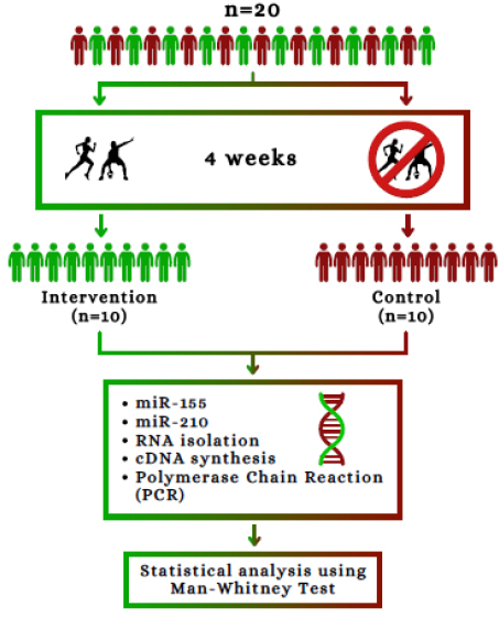
Study design of the intervention study where n=20.
| Week | Frequency (sessions/week) | Duration (min/session) | Intensity (% HRmax) | Total Weekly Volume (min) |
|---|---|---|---|---|
| 1 | 3 | 60 | 65% | 180 |
| 2 | 3-4 | 75 | 70% | 225-300 |
| 3 | 3-4 | 90 | 70% | 270-360 |
| 4 | 3-4 | 100 | 75% | 300-400 |
2.2. Sample
Those who fulfilled the following criteria were considered qualified: aged 25-40 and in good health. The exclusion criteria were as follows: (1) a history of serious mental, neurological, or cardiovascular disorders; (2) pregnancy; (3) muscle pain; and (4) usage of analgesics. For the duration of four weeks, participants were told to maintain their usual dietary patterns and lifestyles. No specific limit on the calories that they could consume was provided, nor was there any recommended type of diet. For each week, they had to fill out self-administered surveys to get qualitative information on their meals, sleep time, and intake of caffeine, alcohol, and nicotine. All the subjects remained consistent throughout the experiment with respect to these factors. The physical activity status was not measured by a validated, standardized physical activity questionnaire, but by self-report screening and weekly self-administered lifestyle questionnaires, which is recognized as a methodological limitation.
2.2.1. Sample Size Rationale
A formal a priori power calculation was not done as this was an exploratory pilot study. To evaluate feasibility, adherence to the supervised exercise protocol, and preliminary changes in circulating miR-155 and miR-210 expression following 4 weeks of endurance training, a sample size of 20 participants (10 endurance training group and 10 age-, sex-, and BMI-matched sedentary control group) was used. Thus, the results should be interpreted with caution and used for hypothesis generation only, and a larger randomized controlled study should be performed to support the effects observed.
2.3. Blood Collection
Venous blood samples (~9 mL) were drawn from the antecubital vein at baseline and at the end of 4-weeks of the intervention. The post-intervention sample was collected from children in the intervention group within 30 minutes of the last supervised training session. In the control group, blood was drawn at both baseline and 4 weeks of continued sedentary behaviour. Hence, the post-intervention miRNA measurement for the exercise group represents the end of the intervention training state, which could include both chronic training adaptation and the acute response to the last exercise bout. In this pilot study, a formal hemolysis index was not evaluated. The blood collection and plasma processing procedures were standardized across all participants to ensure that the minimum pre-analytical variability was achieved. Residual hemolysis may affect measurements of circulating miRNAs, and should be evaluated in future studies.
2.4. miRNA Isolation
Total RNA, including small RNA, was isolated from 200 μL of plasma according to the manufacturer’s instructions provided with the TRI Reagent® kits, with slight modifications to the protocols. In particular, 750 μL of TRI Reagent® was mixed with each sample of plasma, incubated for 5 minutes at room temperature, then 200 μL of chloroform was added; samples were mixed vigorously for 15 seconds and incubated for another 2 to 3 minutes at room temperature. Next, the samples were centrifuged for 15 minutes at 4°C and 12,000 × g. The aqueous phase was collected and transferred to a new RNase-free tube, while RNA was precipitated by adding isopropanol. Afterwards, the RNA was dried under air and re-dissolved in RNase-free water. RNA concentration and purity were measured using a NanoDrop ND-1000 spectrophotometer (Thermo Fisher Scientific, USA). Only samples with A260/A280 ratios between 1.8 and 2.1 and A260/A230 ratios > 1.8 were used for reverse transcription
2.5. Reverse Transcription
The RNA was converted to cDNA, the target MiRNA was discovered, and the miRNA was extended using the PrimeScriptTM RT Kit (#RR037A). The PrimeScript RT Reagent Kit optimizes the reverse transcription step in real-time RT-PCR. It makes use of PrimeScript RTase, which is highly extensible and enables the quick and efficient synthesis of cDNA templates for real-time PCR. The step-experimental method is suitable for high-throughput analysis and is simple to apply. This kit is compatible with a Real-Time PCR reagent called SYBR® Premix.
2.6. Real-Time qRT-PCR
As previously stated, quantitative Real-Time PCR (RT-PCR) was carried out using the TAKARA qPCR Master Mix (2X) Universal Kit. Primers used for this study have been listed in Table 2. U6 (RNU6-1) was used for internal spike-in amplification. The fold change was calculated by dividing the average normalized miRNA expression (2−ΔΔCt) of the test group samples by the average normalized miRNA expression (2−ΔΔCt) of the control group samples. Normalization was performed using a Ct mean of the expressed miRNAs [11].
| miRNA | Sequence (5' → 3') |
|---|---|
| hsa-miR-155-5p | Forward: GCGCGTTTAATGCTAATCGTGATAG |
| Reverse: CAGTGCAGGGTCCGAGGTAT | |
| hsa-miR-210-3p | Forward: GCGCGCTGTGCGTGTGACA |
| Reverse (universal): CAGTGCAGGGTCCGAGGTAT | |
| U6 snRNA (RNU6-1) | Forward: CTCGCTTCGGCAGCACA |
| Reverse: AACGCTTCACGAATTTGCGT |
2.7. Statistical Analysis
Statistical analysis was carried out using IBM SPSS Statistics software. Due to the small sample size of these miRNAs and the non-normal distribution of the data, non-parametric statistical analysis was used. The Wilcoxon signed-rank test was used to compare pre- and post-test scores within groups. Mann-Whitney U test was used to make between-group comparisons (intervention versus control at baseline and at 4 weeks). Spearman’s rank correlation coefficient (ρ) was used to calculate the association between miRNA expression levels and participant/training characteristics. All data are described as mean ± SEM or as medians when appropriate. A p-value of <0.01 was regarded as statistically significant for a two-tailed p-value. The Wilcoxon signed rank test was used for within-group pre- and post-tests. The Mann-Whitney U test was used for between-group comparisons, and Spearman's rank correlation coefficient was used to assess the correlation.
3. RESULTS
3.1. Participants' Attributes
The majority of the participants were college-aged, with nine men and one woman chosen as research volunteers. Only a small fraction followed a strict diet, although the majority ate sensibly. The study group's BMI ranged between 19.3 and 28.6. The exercise program consisted of 3-4 treadmill sessions per week under supervision. Session duration was increased from 60 minutes at week 1 to 100 minutes at week 4. Intensity was increased from 65% to 75% HRmax during the four weeks. The previously reported wide range (3-6 sessions/week; 60-120 min) in the original manuscript reflected participants' self-reported baseline physical activity levels prior to the intervention, rather than the prescribed exercise protocol. The distributions of age, BMI, weekly exercise-session frequency, and exercise-session duration in the intervention group are presented in Fig. (2).
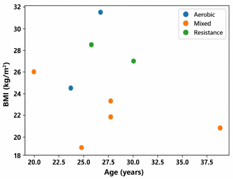
The scatter plots for each value and group mean (horizontal line) with standard deviation (error bar). The variables include age (years), Body Mass Index (BMI; kg/m2), number of exercise sessions per week, and minutes spent on each exercise session for the intervention group (n = 10). The means of the variables, along with their respective standard deviations, include age 31.4 ± 5.2 years, BMI 24.3 ± 2.8 kg/m2, sessions 3.4 ± 0.5 sessions/week, and minutes 81.2 ± 13.4 minutes per session.
3.2. Circulating MicroRNAs
A Wilcoxon signed-rank test was used to determine whether expression of miR-155 was significantly increased in the intervention group compared to the control group, within the intervention group. All intergroup comparisons were made with the Mann-Whitney U test, and all correlation analyses were conducted with Spearman's rank correlation coefficient.
In contrast, miR-210 expression did not change significantly (median pre: 0.729, median post: 0.702; p = 0.73). As shown in Tables 3 and 4, seven out of ten participants (70%) had increased levels of miR-155 post-training compared with pre-training. In contrast, only three individuals, or 30% of the participants, had a slight reduction. The mean value increased from 1.128±0.009 to 1.151±0.010, as revealed by the Wilcoxon signed-rank test at p = 0.008. However, all participants demonstrated only a small reduction in serum miR-210 from pre- to post-training (Tables 3 and 4). Nonetheless, the decrease was very minimal, with an average change of -0.011, which is less than 2% of the baseline level. Moreover, there was no statistical significance based on the Wilcoxon signed-rank test (p = 0.73). Thus, the 4 weeks of endurance training failed to alter serum miR-210 significantly. At baseline, neither miR-155 (median intervention: 1.108 vs. control: 1.101; Mann-Whitney U, p = 0.82) nor miR-210 (median intervention: 0.729 vs. control: 0.735; p = 0.91) showed any significant changes between the intervention group (n = 10) and the control group (n = 10). Following four weeks of intervention, the expression level of miR-155 was significantly increased compared to the control group (median 1.165 vs. 1.104; p = 0.009). On the other hand, there was no significant difference in the expression level of miR-210 between the two groups (median 0.702 vs. 0.718; p = 0.67). (Fig. 3, Table 3).
| Participant | miR-155 (pre) | miR-155 (post) | Fold Change | miR-210 (pre) | miR-210 (post) | Fold Change |
|---|---|---|---|---|---|---|
| 1 | 1.102 | 1.135 | +0.033 | 0.681 | 0.672 | -0.009 |
| 2 | 1.095 | 1.121 | +0.026 | 0.719 | 0.712 | -0.007 |
| 3 | 1.138 | 1.155 | +0.017 | 0.761 | 0.753 | -0.008 |
| 4 | 1.122 | 1.139 | +0.017 | 0.733 | 0.724 | -0.009 |
| 5 | 1.105 | 1.124 | +0.019 | 0.706 | 0.698 | -0.008 |
| 6 | 1.158 | 1.192 | +0.034 | 0.831 | 0.819 | -0.012 |
| 7 | 1.198 | 1.235 | +0.037 | 1.031 | 1.017 | -0.014 |
| 8 | 1.131 | 1.152 | +0.021 | 1.052 | 1.036 | -0.016 |
| 9 | 1.107 | 1.118 | +0.011 | 1.118 | 1.102 | -0.016 |
| 10 | 1.128 | 1.143 | +0.015 | 1.227 | 1.213 | -0.014 |
| Mean ± SEM | 1.128 ± 0.009 | 1.151 ± 0.010 | +0.023 | 0.886 ± 0.062 | 0.875 ± 0.061 | -0.011 |
| Group | miRNA | Pre-training | Post-training | p-value* | Direction |
|---|---|---|---|---|---|
| Intervention | miR-155 | 1.128 ± 0.009 | 1.151 ± 0.010 | 0.008 | ↑ |
| Intervention | miR-210 | 0.886 ± 0.062 | 0.875 ± 0.061 | 0.73 | No change |
| Control | miR-155 | 1.125 ± 0.011 | 1.124 ± 0.010 | 0.82 | No change |
| Control | miR-210 | 0.890 ± 0.060 | 0.888 ± 0.059 | 0.91 | No change |
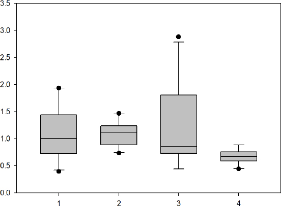
Alterations caused by training in the microRNAs that are in circulation. Expression levels were measured both before and after training. Each point represents the mean of the studied samples. (1) miR-155 pro training and (2) post-training; (3) miR-210 pro training and (4) post-training. The p-values for miR-155 were 0.008, whereas those of miR-210 were 0.73; both were calculated using the Wilcoxon signed-rank test on data obtained before and after training
Due to the non-normal distribution of the data, Spearman’s correlation coefficient (ρ) was used. The correlations between miRNA expression levels and participant/training characteristics are summarized in Table 5. The only association that came close to statistical significance was the one between miR-155 and overall training time (ρ = 0.495, p = 0.021), albeit it did not reach the pre-specified threshold of p < 0.01.
| Variable Pair | Spearman’s ρ | p-value |
|---|---|---|
| miR-155 vs. total training time (hours) | 0.495 | 0.021 |
| miR-155 vs. session frequency (sessions/week) | 0.297 | 0.185 |
| miR-155 vs. age | -0.366 | 0.098 |
| miR-210 vs. age | -0.117 | 0.612 |
| miR-210 vs. session frequency | -0.256 | 0.258 |
| miR-210 vs. total training time | -0.089 | 0.700 |
4. DISCUSSION
The goal of our study was to perceive how hard training affected the levels of particular circulating extracellular microRNAs in plasma and how they related to known physical activity. Our results provide insights into fluctuations in the levels of certain microRNAs, such as miR-155 and miR-210, over the course of a 4-week training cycle. Following the intense training session, we observed a notable increase in miR-155 levels, consistent with the findings of Krammer et al. [10], who showed that exercise training considerably raised blood levels of miR-155. There was no discernible change in the serum levels of miR-210 in ten participants following exercise. While miR-146a expression was sensitive to exercise at a certain threshold but did not exhibit dosage dependency, Ramos et al. (2018) [12] documented that miR-21 and miR-210 expressions were not responsive to exercise. Nevertheless, miR-210 and age (r=-0.117), session frequency (r=-0.256), and miR-155 with age (r=-0.366) were found to be negatively correlated. In recent years, researchers have focused a lot of attention on the function of circulating miRNAs during the exercise adaptation process in an effort to use them as a potential marker to direct disease treatment and athlete training [8, 13, 14]. Circulating blood levels of miR-21, miR-146a, miR-1, miR-210, miR-155, and miR-181 were measured and compared before and after exercise following the 5-kilometer run test. MiR-210, miR-146a, miR-1, miR-181, miR-155, and miR-146a, thus demonstrated a noteworthy rise following the 5-km run test. The two groups did not differ statistically, despite an increase in miR-21 levels. Interestingly, our results did not entirely align with earlier research on exercise. While miR-155 and miR-21 were unaffected by marathon running, miR-1 expression increased in the health training population, indicating a potential function for miR-1 in cardiovascular adaptation mechanisms after endurance exercise [15]. Ramos et al. (2018) [12] reported that although miR-146a expression was dose-independent and responsive to exercise at a certain threshold, miR-210 and miR-21 levels were not. Additionally, exercise has a dose-dependent effect on miR-1 levels as intensity increases [12]. Therefore, Gaál et al. (2017) [6] found that following exercise, miR-181a and miR-21 levels dramatically rose while miR-1 levels stayed constant. We hypothesize that the frequency, duration, intensity, and type of exercise, as well as the subject status and the time point of miRNA monitoring following exercise, are related to the alteration of particular miRNAs during exercise, based on the findings of this study and others.
One essential methodological point to keep in mind is the timing of blood sample collection after the intervention. The circulating miRNA profile observed in the intervention group may reflect chronic training adaptation, as well as the acute miRNA response to the last supervised exercise session. These results should thus not be viewed as strictly resting-state adaptations, but as responses to the exercises performed at the end of the intervention. There are a number of methodological and physiological factors to consider when interpreting the response to exercise in circulating miRNAs. The amount and direction of circulating miRNA alteration may depend on the intensity and duration of exercise, and previous studies demonstrate that there is differential response of some c-miRNAs respond differentially to changes in endurance exercise dose. Training status is important, since sedentary people, recreationally active people, and trained athletes may have varying miRNA baseline values and different training responses to the same exercise. Another important factor is the timing of sampling, as the levels of circulating miRNAs can fluctuate rapidly following exercise and can even partially or completely normalize during recovery, depending on the type of exercise and/or miRNA of interest. Last, but not least, miRNAs in circulation do not have to originate from a single tissue. While a portion of miRNAs are localized in particular tissues such as skeletal muscle, cardiac muscle, endothelial tissue, or immune cells, plasma miRNA signals are a systemic signal, which is subject to tissue release and vesicle uptake in the extracellular space and to clearance mechanisms. Hence, plasma miR-155 levels were increased, and it is important to note that miR-210 was not significantly altered in the present study; however, these should be interpreted with caution as a response to circulating miRNAs and not as expression changes in the tissue of origin. Clinical biochemical markers (Myo, CK-MB, LDH, IL-6, IMA, and troponin) were not measured in the present study. As such, it was not possible to test the direct association between miRNA expression and biochemical markers of inflammation, muscle damage, or myocardial stress. Circulating miRNA profiling should be integrated with other biochemical and physiological measures in future studies to improve understanding of the biological significance of exercise-induced changes in miRNA levels. Acute exercise has been shown to increase CK, LDH, and the N-terminal segment of the BNP precursor (NT-proBNP), but not CK-MB, cardiac troponin T (cTNT), or high-sensitivity C-reactive protein (hs-CRP) [16, 17]. It has been fould that serum levels of LDH, IL-6, AST, Myo, and CK-MB significantly increased after the 5-kilometer running training, but Inferior Mesenteric Artery (IMA) levels were decreased. These results imply that young, healthy participants clearly adapt to exercise. Additionally, exercise increases IL-6 release in muscles and initiates anti-inflammatory responses by promoting IL-10 secretion and inhibiting IL-1 release [18, 19]. Nonetheless, most studies show that IMA can increase significantly in cases of severe myocardial ischemia, such as coronary heart disease [20]. MiR-146a suppresses inflammation and myocardial injury by inhibiting the TLR4/NF-κB signaling pathway, while miR-155, miR-1, and miR-181 control target genes to suppress inflammation in myocardial hypoxia [20, 21]. It is important to note that this investigation did not include an evaluation of any of the clinical biochemical markers (AST, LDH, CK-MB, IL-6, IMA, troponin, etc.). Any information relating to these markers is provided solely by references to scientific literature, as these data are used to give some background on the biological processes involved in miRNA regulation. Moreover, most published research articles indicate that IAM is an essential inflammatory marker for most diseases, such as rheumatoid arthritis, cardiovascular disease, and transient ischemic attacks [22, 23]. During exercise training, miRNAs may control IL-6 release, inhibiting the inflammatory response and reducing IAM levels. In our present study, however, miR-146a, miR-155, and miR-210 were positively correlated with Myo, CK-MB, and LDH, respectively. The findings suggest that other miRNAs than those mentioned above can regulate the markers of inflammation and heart damage by controlling the inflammatory condition of young subjects. Further research is needed to determine whether these miRNAs have a direct effect on the above metabolic parameters. The sole limitation of this study is the fact that miRNA expression was measured in a fairly small population. Other limitations involve our individuals' demographic profile, including gender, as most participants were male. Despite matching for demographic characteristics, the absence of randomization and selection of participants into experimental or control groups makes it possible for selection bias to be present in this study. Finally, there may be unmeasured confounding because dietary consumption was documented by self-report rather than controlled. To validate our results and investigate sex-specific miRNAs, more research with larger, sex-balanced populations and randomized controlled methods is required [24]. The exercise-induced regulation of miRNAs such as miR-155 and miR-210 has the potential to serve as a valuable complement to drug or medicinal chemistry-driven interventions for disease management by leveraging the body's endogenous regulatory mechanisms. Exercise has been shown to regulate the miRNA expression profile in a context-dependent and tissue-specific manner, thereby targeting key pathways responsible for inflammation, immunity, angiogenesis, and oxidative stress. For example, exercise-induced downregulation of miR-155 could inhibit chronic inflammation and restore immune homeostasis, complementing anti-inflammatory medication and possibly reducing its dosage and side effects. Similarly, exercise-induced upregulation of miR-210 could enhance angiogenesis, improve mitochondrial function, and protect endothelial cells against oxidative stress, thereby enhancing the therapeutic effects of pro-angiogenic drugs or cardioprotective agents. Unlike pharmacological therapies that tend to act on isolated molecular targets, exercise might have systemic actions, influencing various signaling cascades and miRNA networks simultaneously. This holistic regulation is synergistic in advantage, as exercise can enhance drug effectiveness, decrease side effects, and improve patient outcomes overall. Hence, the integration of exercise-induced miRNA regulation with conventional therapies is a comprehensive and efficient approach towards treating complex diseases such as cardiovascular diseases, cancer, neurodegeneration, and metabolic disorders.
5. LIMITATIONS OF THE STUDY
The results of this study have to be interpreted with regard to a number of limitations. First, the sample size was small (as with any pilot study), so the results should be taken as preliminary. Second, the sample was not sex balanced, with more males than females, which might restrict the generalizability of the results to females and also preclude examination of sex-specific miRNA responses. Third, the study was a non-randomized study; age, sex, and BMI were matched across groups, but there is still a possibility of selection bias. Fourth, volunteer bias may have been present because the volunteers who agreed to participate in the endurance training programme may have differed from those who did not, in both motivation to participate and health awareness and/or lifestyle stability. Adherence to the programme was assessed using attendance records and heart rate data, confirming that all participants attended at least 90% of the sessions. However, minor differences in adherence, exercise effort, recovery, and physical activity outside the supervised training sessions may have contributed to interindividual variability in the miRNA response. Furthermore, the postintervention blood sample was collected within 30 minutes after the final exercise session, which makes it difficult to distinguish acute exercise from chronic training adaptations. Lastly, physical activity and lifestyle habits were self-reported, but not measured by a validated and standardized physical activity questionnaire, which may introduce recall bias or residual confounding. Additionally, there was no formal hemolysis index measured, and the potential impact of the residual hemolysis on circulating miRNA levels has not been ruled out.
CONCLUSION
Based on our data and within the limitations of this pilot study (n = 10 per group), we draw the following conservative, data-driven conclusions: a 4-week supervised endurance training program in previously inactive, healthy adults resulted in a statistically significant increase in circulating miR-155 (Wilcoxon signed-rank test, p = 0.008), with 7 out of 10 participants showing elevated post-training levels; however, the absolute increase was modest (~2%), and we did not measure functional outcomes (e.g., inflammatory cytokines), so the biological significance remains unknown. The same intervention did not yield a statistically noteworthy alteration in the expression level of miR-210 (p = 0.73), in spite of a consistent small numerical decline across all individuals. Total training duration and miR-155 change showed a somewhat favorable association (Spearman's ρ = 0.495, p = 0.021), but it fell short of our predetermined criterion of p < 0.01, and no other correlations were significant. Inter-individual variability in miR-155 response was notable (fold change range: 0.98-1.15), suggesting possible responder/non-responder differences even to a standardized program. To our knowledge, no previous study has specifically examined the combination of miR-155 and miR-210 in response to a 4-week endurance training program in previously inactive, healthy adults with a matched non-intervention control group. This specific combination of miRNAs, population (inactive adults), intervention duration (4 weeks), and controlled design limits direct comparisons with prior work, which has largely focused on acute exercise, athletic populations, or different miRNA panels
AUTHORS’ CONTRIBUTIONS
The authors confirm contribution to the paper as follows: M.A-M.: Study conception and design; F.M.: Data collection; M.A-M.: Analysis and interpretation of results; M.A-M.: Draft manuscript; E.E., M.G.N.A., L.H.: Visualization and validation. All authors reviewed the results and approved the final version of the manuscript.
LIST OF ABBREVIATIONS
| HRmax | = maximal Heart Rate |
| HUVECs | = Human Umbilical Vein Endothelial Cells |
| MSCs-EVs | = Mesenchymal Stem Cell-derived Extracellular Vesicles |
| IMA | = Inferior Mesenteric Artery |
ETHICS APPROVAL AND CONSENT TO PARTICIPATE
The study was approved by the institutional ethics committee of Sohar University, Oman with the decision number UEB2024-028.
HUMAN AND ANIMAL RIGHTS
All human research procedures followed were in accordance with the ethical standards of the committee responsible for human experimentation (institutional and national), and with the Helsinki Declaration of 1975, as revised in 2013.
CONSENT FOR PUBLICATION
Before enrolling in the training program, all individuals provided verbal and written informed consent.
AVAILABILITY OF DATA AND MATERIALS
The data and supportive information is available within the article.
ACKNOWLEDGEMENTS
Declared none.